Osteoarthritis of the hip and degenerative diseases-degenerative disease that includes the destruction of hyaline cartilage. The disease develops gradually, accompanied by an armbar syndrome, and a reduction in the number of entries. In the absence of any medical intervention in the early stages of arthritis, a few years later, it occurs to atrophy. femoral muscle. The leg is shortened, and the fusion-joint crack resulting in partial or complete paralysis of the hips. The causes of the pathology, if you make it prior to the injury, curvature of the spine, diseases of the musculoskeletal system.

Osteoarthritis is generally found in patients are middle-aged and older adults. The diagnosis is explained, based on the findings of the research tools that are available for the x — ray, magnetic resonance imaging, computed tomography, arthroscopy. The treatment of the pathology of the 1-and 2-degree-of-severity-conservative. When it is detected that an ankylosis, or failure of medical therapy is performed, an operation (arthrodesis, arthroplasty).
The mechanism of the development of the disease
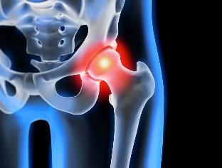
The articulation of the hip to form two bones — the iliac and buttock. Lower division and lower abdomen, represented by its body, which is involved in the plexus: the femoral, which is at the top of the department is in the cup. During the movement of joint without the custom property, and the buttocks, the head can move freely. This is the "rotation" system of articulation that allows you to bend, stretch, twist, and promotes abstraction, and to bring to the thigh. The good sliding of the structures in the joint ensures a stretchy, elastic, hyaline cartilage, the lining of a duct, and the head of the femur. Its main function is the redistribution of the charges in the motion, a notice of the rapid wear and tear of the tissues to the bone.

Under the influence of internal or external factors disturbed trophic cartilage. It doesn't have a circulatory system — the nutrients of a fabric, it provides the synovial fluid. When you win, she's grown up, it becomes a sticky paste. Came up for the deficiency of nutrients, it causes a drying out of the surface of hyaline cartilage. It is covered with cracks, which leads to a constant micro-damage to tissues during the flexion and extension of the fine from the hip. The cartilage of the antarctic are losing their particular properties. To "adapt" to the increase of the pressure, to deform with the bones. And in the midst of the deterioration of the metabolism in the tissues, enhance the destructive-degenerative changes.
The causes and factors that may cause the
Idiopathic or primary osteoarthritis develops without any reason. It is believed that the destruction of the articular cartilage tissue due to aging of the organism, slowing down the recovery process and reduce the production of new collagen and other compounds that are required for the complete regeneration of the structures of the pelvis. Secondary osteoarthritis-it occurs at the bottom of this in the body in a pathological state. The most common reason for secondary disease include the following:
- prior to the injury, is damage to the tendon apparatus, for breaks in the muscles, to a complete separation of the bone marrow foundation, fractures, dislocations;
- a violation of the development of mating, birth and dysplastic disease;
- the pathology of the autoimmune disease — rheumatoid arthritis, reactive arthritis, psoriatic arthritis, systemic lupus erythematosus, systemic;
- it is non-specific and inflammatory disease, for example, purulent arthritis;
- sexually transmitted infections — gonorrhea, syphilis, brucellosis, ureaplasmosis, trichomoniasis, tuberculosis, osteomyelitis, swelling of the brain;
- a violation of the functioning of the endocrine system;
- degenerative diseases degenerative disease — osteochondropathy head of the femur;
- hypermobility joints, due to the release of "over-stretch" of the collagen, causing an excess of mobility, weakness of the joints.
As well as the root cause of the development of osteoarthritis, and can become a hemarthrosis (blood in the cavity of the hip), and the inflammatory factors that are related to the violation of hematopoietic. A pre-requisite for the occurrence of this disorder are excessive weight, excessive exercise, sedentary lifestyle. Its further development leads to an incorrect organisation of the sport, the training, the deficit in the diet of foods with a high content of vitamin d, giraud and also vitamins that are soluble in water. The post-operative osteoarthritis-it occurs a few years later, after the surgery, especially if it is accompanied by an excision of a large amount of the body's tissues. Osteoarthritis of the hip, it can't be inherited. But while some characteristics are innate (disorder of the metabolism and the structure of the skeleton), the probability of her developing it is greatly increased.
The symptoms of
The main symptoms of osteoarthritis of the hip, pain when walking, in the region of the hip joint and the knee joint. A man, suffering from stiffness of movement, stiffness, especially in the morning. In order to stabilize the joint, and the patient begins to limp, to change to a different gear. Over time, due to muscle atrophy, and the deformity of the joint of a limb is visibly reduced. Another feature of the pathology of the limit of the displacement of the hips. For example, the difficulties that arise when one tries to sit down on a stool, spreading her legs out to the sides.

Even the "cast" of the ARTHRITIS it can be cured at home. And don't forget, a once-a-day to film this...
For osteoarthritis the first level of severity is a characteristic of the periodicals of the pain occurs right after an intense physical effort. They are located in the vicinity of the joint, and then disappear after a long period of time for the holidays.
When you read the second-degree connection to the demonstration starting point of the pain syndrome, is increased. The uneasy feelings occur even in the resting state, it has spread over the thighs and the groin are strengthened with the increase in the severity of, or improve the training of the motor activity. Eliminate the pain in the hip, and the man begins a barely noticeable limp. Notes limitation of motion in the joint, especially if the warranty, and internal rotation of the hip joint.
With the win, the third kind, which is characterised by constant severe pain. When the movement is in trouble, so when you walk in, the man is forced to use a cane or crutches. Due to the weakness of output, femoral muscle occurs, the displacement of the bone of the pelvis in the frontal plane. To compensate for what has happened in a shortened foot, the patient will move and the slope in the direction of the damaged members. This results in a strong displacement of the center of gravity, and the increase of the loads on the joint. At this stage in the evolution of arthritis, marked ankylosis of joint.
| The degree to which | The radiographic signs of |
| The first | The changes are expressed not sharply. The joints crack a little, equally narrowed to the lack of damage to the surface of the thigh. To use the external or the internal rim of the acetabulum is seen a small proliferation of bone |
| The second | The height of the slit joint is reduced significantly, due to the uneven fit. The bones of the head, the hips pushed up, distorted, enlarged, and their contour becomes irregular. Bone tumors are formed on the inner surface and the outer edges of the fossa to the articular |
| The third | Occurs in a total or partial joint fusion crack. The head of the femur is strongly expanded. Multiple bone tumors that are located in all areas of the acetabulum |
The diagnosis
To make a diagnosis, the physician takes into consideration both the clinical manifestations of the disease, the clinical history, the results of the visual inspection of the patient and of the instruments of the research. The most informative x-ray. With your help, is to assess the state of the hip joint, is defined by the phase of the current, and the degree of damage of the cartilage tissue and, in some cases, the cause of development. If a cervical-diaphyseal site, increased vertluzhnoj depression, bezel, and flat-topped, with high probability, it can be assumed dysplastic changes inherent to the hinge. The disease Pertusa indicate damaged on the way to the bone of the thigh. The x-ray allows you to vyvit of post-traumatic arthrosis, in spite of the absence of a previous history of disease of injury. They are also used for other diagnostic methods:
- The ct scan helps in detecting the expansion of the edges of the plates to the bone, set the bone spurs;
- A magnetic resonance imaging performed for assessment of the status of the connective tissue structures, and the degree of their involvement in the pathological process.
If necessary, the inner surface of the joint be examined with the arthroscopic tools. The differential diagnosis is carried out, to exclude, to gonarthrosis, pain, sacral, or degenerative disc disease. Pain in osteoarthritis may disguise the clinical manifestations of radicular syndrome, caused by the violation, or to an inflammation of the optic nerve. To exclude neurogenic pathology generally, it is possible by means of a series of test cases. Osteoarthritis of the hip, it is necessarily differentiated, higher-bursitis of the hip joint disease, Spondylitis, reactive arthritis. To the exclusion of autoimmune disorders, are conducted biochemical studies of blood and synovial fluid.
Surgery
When the inefficiency of the conservative therapy of an il diagnosis of a complicated disease is carried out in the operation. To recover the cartilage tissue in the joint, damaged, read, no operation, fitting of artificial limbs is impossible, but with the right approach, to treatment, to comply with any of the provisions of the medical, which is run by a correct life-style lessons and physical therapy sessions because of the regular, as well as massage, reception of vitamins and the proper nutrition, you can stop the process of the defeat and destruction of the cartilage and the joints of the hip joint.


